Be proactive. Early detection saves lives. No radiation, pain, compression or blood draw. Ideal for those with implants.
Scans the breast/chest for inflammation, lymph node and hormone activity and digestion issues.
PARTIAL BODY IMAGING - $350
This scan images from the head to the pelvic bone, thyroid, periodontal, carotid artery, full spine, the small and large intestine, gallbladder, liver, kidneys, bladder, uterus, ovaries (F), prostate gland (M).
FULL BODY IMAGING - $525
The Full Body Scan is perfect for looking at the big picture. This scan images from the bottom of the feet to the top of the head. Includes all the imaging of the Partial Body Imaging plus arms, legs, hands, and feet.
FOLLOW UP IMAGING - $175
Necessary for comparative analysis screening. This imaging test will target the sites of concern and is a wonderful comprehensive to your baseline imaging.
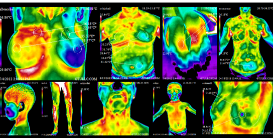
WHAT IS THERMAL IMAGING?
Thermal imaging, FDA approved in 1982, is an unsurpassed, safe and noninvasive disease-screening technique that can detect signs of tumors up to ten years earlier than is possible using other tests. Inflammation and other conditions can immediately be detected as well such as periodontal disease or infection (that may go unnoticed by x-ray) which is incredibly important to detect early when it comes to heart health and breast health.
Clinical Thermography, also known as Digital Infrared Thermal Imaging is a simple test of physiology that relies on the sympathetic nerve control of skin blood flow and the ability of the sympathetic nervous system to respond to (and react to) pain, pathology, injury or dysfunction anywhere in the body. While other diagnostics, such as ultrasound, radiography and mammography show the body’s structure or anatomy, DITI is unique in that it shows physiological and metabolic processes.
Thermal imaging is an extremely accurate heat sensitive imaging device that uses definitive variation of colors to represent actual temperature variations for that specific region of the body. The primary view held by thermographers & doctors is that our body should have corresponding heat patterns for both sides of the body mid line on both sides. A standard deviation that is considered significant is a variation of 0.3 C degree or greater.
How skin temperature renders a health status of the body is shown below:
In fetal development, the skin, brain, and spinal cord originate from the same cellular tissue. Skin temperature control is directly “hotwired” to the brain and spinal cord so as to constantly regulate the body core temperature and thus maintain the health of the entire system.
The skin temperature is regulated by that part of the brain that also controls the blood flow to every muscle, organ, and gland.
Any changes in the internal issues and environment of the body tend to produce a reflexive and corresponding change in skin temperatures that show very graphically in a thermographic image. Heat imaging gives visual points of reference regarding the body’s ability to work correctly. We thus can see the functional, physiological, and biological pattern of the blood flow within the body. Heat patterns, either higher or lower, tend to take on the color patterns of that part of a body being affected by an outside influence that is different from the norm
Our focus on breast health is related to breast cancer precursors–those heat markers and areas that no x-ray or mammogram could ever show, such as:
(1) Chronic (prolonged) inflammation
(2) Chronic lymphatic congestion (the systems that feed breast tissues and carry waste away)
(3) Estrogen inflammatory influences (frequently, blood test results show normal prognosis)
Thermography is not considered an independent (solely used) diagnostic tool. There are other considerations such as breast lesions that must be considered beyond the scope of thermography. But on the other hand, common disorders like strains are not even noticed by X-ray, MRI, or CT scan. Neither can issues such as sciatica or neuropathies such as Reflex Sympathetic Dystrophy (RSD) be detected with any method besides thermography. The limitations of x-ray and the numerous other scan methods are enhanced with thermography. It is realized that only a biopsy can give a definitive diagnosis of cancers.
Dr. Gregory Melvin
Dr. Melvin - Doctor of Chiropractic
Board Certified Thermal Reading Doctor for 20+ Years
Dr. Melvin has 35 years of chiropractic experience, 20 years of reading and interpretations of thermal imaging scans and 300 hours of radiology. He is a noted speaker who teaches and certifies doctors and technicians in thermal imaging. He has trained offices throughout the US and continues to educate himself everyday in analyzing his patient scans to help them have a better understanding of their health and health options.
Thermal imaging has been able to help in the early detection of inflammatory breast cancer which frequently affects women between the ages of 20 and 40. Thermal imaging can screen for the earliest stages of cancer in ALL women, all age groups, and is often preferred to mammograms since this technology has NO RADIATION and NO BODY CONTACT, and is done in a matter of minutes while you wait. It is currently recognized as one of the earliest methods of screening for breast disease. The American Heart Association suggests thermal imaging as an initial screening for heart disease.
The reality is ALL women face the threat of breast cancer. Current statistics show that one-in-eight American women will be diagnosed with the disease in her lifetime.
For over thirty years we have been told that an annual mammography examination is the key to long-term survival from breast cancer. Yet, after thirty years there has been no significant improvement in mortality.
FAQ's ON THERMOGRAPHY
What are the benefits of Thermal Imaging?
Thermography is a non-invasive diagnostic tool with scientifically valid, reproducible results.
Thermography is an FDA approved adjunctive diagnostic device so the results can be trusted.
Thermography can detect dysfunction even when conventional blood tests and radiographictests were negative.
Does insurance cover thermal imaging?
All insurance carriers are different. Currently Total Thermal Imaging does not accept insurance, however they can provide you with a Superbill for you to submit to your insurance company.
HSA or FSA accounts are accepted as a viable method for paying for your scan.
Why get tested with thermal imaging?
Thermal imaging has been an available medical test for more than 50 years, and its nonmedical applications pre-date that by a decade.
Despite controversy in its early days, this tool has withstood the test of time and has become the mainstay in many diagnostic endeavors. The technology in modern day thermography as well as properly training technicians has improved tremendously over the years.
With the increasing technological advances of thermal imaging cameras and the newest and best software available, WHY NOT BE TESTED WITH THERMOGRAPHY?
It locates dysfunction with precision going right to the source of heat increased blood supply and enables your doctor to develop a more exact treatment plan. Clearly shows future disease tendencies of those in the process of development.
How does thermal imaging work?
Thermography registers skin-surface temperature from 112 different points on the body.
Thermography displays an image yielding a scan of 25 organs, tissues, or systems and their function.
While X-rays give a structural view, thermography gives us a functional perspective based on physiology and cold stress response.
By computer analysis of the skin temperature patterns, the doctor gains a direct index of the metabolic activity in various parts of the body.
The thermography scan will also show inflamed, degenerated, or overactive organ or tissue processes.
Who should be tested with thermal imaging?
There is no one who shouldn’t be tested. It is a safe procedure for people of all ages. Thermography offers reproducible and scientifically valid information that can be crucial to the development and tracking of a successful treatment strategy.
HISTORY OF THERMOGRAPHY
Thermography has a long history. Breast thermography was discovered in 1956 in Montreal, Canada and rapidly became popular throughout the world. It was FDA cleared and then certified by the American Medical Association as an adjunctive screening procedure for breast cancer in 1982. Recent advancements in technology have allowed us to perform even more accurate exams. A 2008 Study published in the American Journal of Surgery, performed at New York Presbyterian Hospital Cornell showed a 97% sensitivity in discriminating cancer compared to biopsy.
The Biomedical Handbook says:
“In 1982 FDA approved Thermography as an adjunctive Breast screening procedure. Breast Thermography has the ability to detect the first signs of a tumor that may be forming up to 10 years before other procedures can detect it. The greatest evidence supporting the underlying principle of thermal imaging regarding cancerous tumors surrounds the well-documented recruitment of existing vascularity and diagnosis. Conclusion: Thermography has the ability to signal that pre-cursors, such as lymph congestion, hormone imbalance and inflammation may be forming, and set up an ‘alarm’ as much as 10 years before other procedures can recognize them.”
Although medical thermography has been around for several decades, it has not received the attention and credit it deserves from the medical establishment. However, there is evidence of more patients becoming aware of this amazing technology and it’s not unusual that the patient is the one who ends up informing their physician about thermography.
PRE-SCAN INSTRUCTIONS:
YOU MUST WAIT AT LEAST THREE (3) MONTHS AFTER MAJOR BREAST SURGERY, COMPLETION OF CHEMOTHERAPY, OR RADIATION BEFORE A THERMAL EXAM
YOU MUST WAIT AT LEAST ONE (1) MONTH AFTER A BIOPSY OR MINOR SURGERY AVOID TANNING OR SUNBURN AT LEAST ONE WEEK PRIOR TO APPOINTMENT
24 HRS PRIOR TO APPT FOR WHS/MHS/BREAST STUDY
NO SHAVING
ON THE DAY OF YOUR EXAM
DO NOT USE LOTIONS, POWDERS, OR DEODORANTS
AVOID TAKING ANY MEDICATIONS OR SUPPLEMENTS
PLEASE WEAR LOOSE FITTED CLOTHING
IF HAVING A FULL BODY SCAN PLEASE WEAR OR BRING SOCKS WITH YOU TO THE EXAM
12 HRS PRIOR TO EVERY APPT
NO SUNBATHING (SUNBURN)
NO EXERCISING
2 HRS PRIOR TO YOUR APPT:
DO NOT APPLY ANY LOTIONS, CREAMS, DEODORANTS OR POWDERS
NO EATING FOOD
DO NOT EAT OR DRINK ANY CAFFIENE PRODUCTS
DO NOT SMOKE OR USE ANY PRODUCTS CONTAINING NICOTINE
AVOID TAKING ANY MEDICATIONS UNTIL AFTER THE EXAM
AVOID ANY TREATMENTS THAT DAY FOR EXAMPLE (CHIROPRACTIC, ULTRASOUND, MASSAGE ETC)
NO MINTS OR GUM CHEWING
1 HR PRIOR TO YOUR APPT:
DO NOT EAT
Please inform your Thermography Technician about any recent skin lesions or bruising, Rosacea or any other skin disorders as it may cause false positives on reports.
**Necessary medications-such as blood pressure or heart medications are permitted-please notify the technician
PROCEDURE:
To receive accurate results, the equilibrating time (the time it takes for your body to acclimate to the room temperature) is 15 minutes and the room temperature will be between 68-72 degrees. You will be asked to disrobe according to the type of scan chosen and the imaging will take approximately 20 – 45 minutes.
TEST RESULTS:
Reports are generally received within 7 to 10 BUSINESS days after your initial visit. Phone and/or Skype consultations with Dr. Melvin to go over your results are available for an additional fee.